Surface Anatomy
The liver is covered with visceral peritoneum over most of its surface and is suspended by several ligaments.

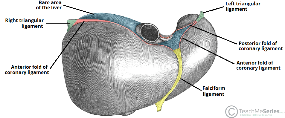
The falciform ligament (with the ligamentum teres at its free margin) is the embryonic remnant of the ductus venosus (umbilical vein) of the umbilical cord. It is a peritoneal structure that courses between the anatomical left and right lobes of the liver and the anterior abdominal wall.
The posterior layers of the coronary ligaments converge to form the lesser omentum, which passes from the visceral surface of the liver to the lesser curvature of the stomach (hepatogastric ligament) and the first part of the duodenum (hepatoduodenal ligament).